
Radiology
Radiology at Tirupati Trauma Care Centre & Hospital
The Radiology Department at Tirupati Trauma Care Centre & Hospital (TTCH) provides diagnostic imaging services that support the evaluation, diagnosis and management of a wide range of medical and surgical conditions.
Radiology plays an important role in trauma care, orthopaedics, neurosurgery, spine surgery, plastic and reconstructive surgery, diabetic foot care and other specialties, helping doctors identify injuries, abnormalities and changes within the body.
At TTCH, the appropriate imaging investigation is selected based on the patient's symptoms, clinical history, suspected condition and area being examined. Our radiology services include X-ray, Ultrasound, CT Scan and MRI.
The imaging findings help treating specialists make informed decisions about further investigation, treatment and follow-up.
The goal is to provide reliable diagnostic imaging and timely clinical information that supports effective patient care.
Radiology Services
1. X-Ray
X-ray is a commonly used diagnostic imaging technique that uses a small amount of ionising radiation to produce images of structures inside the body.
X-rays are particularly useful for evaluating bones and joints and are frequently used in trauma and orthopaedic care.
X-Ray may help evaluate:
Fractures
Dislocations
Bone alignment
Joint abnormalities
Chest conditions
Certain foreign bodies
Postoperative bone and implant assessment
X-ray is often one of the first imaging investigations recommended when a patient presents with a suspected fracture or other bone related injury.
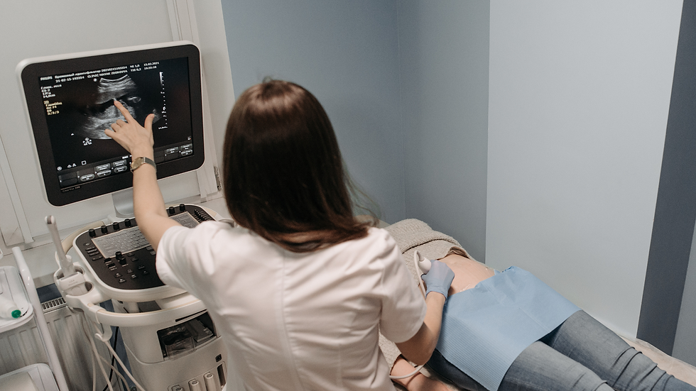
2. Ultrasound
Ultrasound uses high frequency sound waves to produce real time images of structures inside the body. It does not use ionising radiation.
Ultrasound can be used to evaluate selected abdominal organs, soft tissues, muscles, tendons, fluid collections and other structures.
Ultrasound may help evaluate:
Abdominal conditions
Soft tissue abnormalities
Muscle and tendon problems
Fluid collections
Selected paediatric conditions
Certain organ abnormalities
The specific type of ultrasound examination depends on the patient's symptoms and the area being investigated.
3. CT Scan
Computed Tomography (CT) uses X-rays and computer processing to create detailed cross sectional images of the body.
CT can provide more detailed information than a conventional X-ray and can be particularly useful in emergency and trauma care.
CT Scan may help evaluate:
Head and brain injuries
Skull fractures
Complex fractures
Spine injuries
Facial and jaw injuries
Chest injuries
Abdominal trauma
Internal bleeding
Selected tumours and other conditions
CT may be performed with or without contrast depending on the clinical indication.
3. MRI
Magnetic Resonance Imaging (MRI) uses a strong magnetic field and radio waves to produce detailed images of internal structures. MRI does not use ionising radiation.
MRI provides excellent visualisation of many soft tissues, joints, the brain, spine, spinal cord, muscles, ligaments and nerves.
MRI may help evaluate:
Brain conditions
Spine and spinal cord disorders
Disc problems
Ligament and tendon injuries
Joint conditions
Muscle and soft tissue abnormalities
Nerve related conditions
Selected tumours
Before an MRI, patients should inform the medical team about implanted devices, metal fragments, previous surgeries and other relevant medical information

Frequently Asked Questions
What imaging services are available at TTCH?
The Radiology Department at TTCH provides X-ray, Ultrasound, CT Scan and MRI services based on clinical requirements.
Which scan is best for my condition?
There is no single imaging test that is best for every condition. The appropriate investigation depends on your symptoms, medical history, suspected diagnosis and the body part being examined.
What is the difference between an X-ray and a CT scan?
Both use X-rays, but a CT scan uses computer processing to create detailed cross sectional images of the body. CT can provide significantly more information in many complex injuries and trauma cases.
Does an MRI use radiation?
No. MRI uses a magnetic field and radio waves rather than ionising radiation.
Is ultrasound safe?
Ultrasound does not use ionising radiation and is widely used for diagnostic imaging. Your doctor will recommend the appropriate examination based on your clinical needs.
Can an X-ray detect a fracture?
Yes. X-rays are commonly used to identify fractures and assess bone alignment. CT or MRI may sometimes be recommended when additional information is required.
When is a CT scan used after an accident?
CT may be recommended when doctors need detailed assessment of injuries such as head trauma, complex fractures, facial injuries, spine injuries or internal injuries.
When is an MRI recommended?
MRI may be recommended when detailed assessment of soft tissues, the brain, spine, spinal cord, joints, muscles, ligaments or nerves is required.
Do I need preparation before radiology tests?
Preparation depends on the type of examination. Some CT or ultrasound examinations may require specific preparation, while X-rays and many MRI examinations may not. Patients should follow the instructions provided before their scheduled investigation.
Why is radiology important in trauma and orthopaedic care?
Imaging helps doctors identify and assess injuries such as fractures, dislocations, head injuries, spine injuries and complex soft tissue conditions, supporting decisions about treatment and surgery.
What imaging services are available at TTCH?
The Radiology Department at TTCH provides X-ray, Ultrasound, CT Scan and MRI services based on clinical requirements.
Which scan is best for my condition?
There is no single imaging test that is best for every condition. The appropriate investigation depends on your symptoms, medical history, suspected diagnosis and the body part being examined.
What is the difference between an X-ray and a CT scan?
Both use X-rays, but a CT scan uses computer processing to create detailed cross sectional images of the body. CT can provide significantly more information in many complex injuries and trauma cases.
Does an MRI use radiation?
No. MRI uses a magnetic field and radio waves rather than ionising radiation.
Is ultrasound safe?
Ultrasound does not use ionising radiation and is widely used for diagnostic imaging. Your doctor will recommend the appropriate examination based on your clinical needs.
Can an X-ray detect a fracture?
Yes. X-rays are commonly used to identify fractures and assess bone alignment. CT or MRI may sometimes be recommended when additional information is required.
When is a CT scan used after an accident?
CT may be recommended when doctors need detailed assessment of injuries such as head trauma, complex fractures, facial injuries, spine injuries or internal injuries.
When is an MRI recommended?
MRI may be recommended when detailed assessment of soft tissues, the brain, spine, spinal cord, joints, muscles, ligaments or nerves is required.
Do I need preparation before radiology tests?
Preparation depends on the type of examination. Some CT or ultrasound examinations may require specific preparation, while X-rays and many MRI examinations may not. Patients should follow the instructions provided before their scheduled investigation.
Why is radiology important in trauma and orthopaedic care?
Imaging helps doctors identify and assess injuries such as fractures, dislocations, head injuries, spine injuries and complex soft tissue conditions, supporting decisions about treatment and surgery.
Ultrasound
.png)

CT scan
.png)
X-Ray